Review


murine renal carcinoma cell line renca  (ATCC)


Bioz Verified Symbol ATCC is a verified supplier
Bioz Manufacturer Symbol ATCC manufactures this product  
  • Logo
  • About
  • News
  • Press Release
  • Team
  • Advisors
  • Partners
  • Contact
  • Bioz Stars
  • Bioz vStars
  • 96

    Structured Review

    ATCC murine renal carcinoma cell line renca
    L. bacterium protects against ccRCC tumorigenesis through its supernatant (A) Experimental design: After gut microbiota depletion with ATB, <t>RENCA-bearing</t> mice were gavaged with L. bacterium or P. copri 5 times/week ( n = 6/group). (B and C) Tumor growth kinetics (B) and tumor weights (C) on day 24 in mice receiving L. bacterium or P. copri ( n = 6/group). (D) 16S rRNA sequencing of fecal samples from control and L. bacterium -treated mice on days 12 and 24. (E) Experimental design: RENCA-bearing mice pretreated with ATB were gavaged with L. bacterium with or without amoxicillin in drinking water ( n = 9/group). (F and G) Tumor growth curves (F) and tumor weights (G) on day 24 in the indicated groups ( n = 9/group). (H–J) Effects of L. bacterium SN on 786-O <t>and</t> <t>Caki-1</t> cells: colony formation (H), cell viability via CCK8 assay (I), and wound healing assay (J) with 100× and 10× dilutions of SN. Scale bars: 200 μm. (K) Experimental design: ATB-pretreated RENCA-bearing mice were gavaged with L. bacterium , heat-inactivated L. bacterium , or SN ( n = 9/group). (L and M) Tumor weights in BALB/c (L) and BALB/c-nude mice (M) on day 24 ( n = 9–10/group). Data are shown as mean ± SEM. Statistical significance was determined by two-way ANOVA (B, F, and I), unpaired two-tailed t test (C, G, L, and M), and paired two-tailed t test (J). ∗ p < 0.05, ∗∗ p < 0.01, and ∗∗∗ p < 0.001. See also and .
    Murine Renal Carcinoma Cell Line Renca, supplied by ATCC, used in various techniques. Bioz Stars score: 96/100, based on 538 PubMed citations. ZERO BIAS - scores, article reviews, protocol conditions and more
    https://www.bioz.com/product/murine+renal+carcinoma+cell+line+renca/pmc12629829-369-12-22?v=ATCC
    Average 96 stars, based on 538 article reviews
    murine renal carcinoma cell line renca - by Bioz Stars, 2026-08
    96/100 stars

    Images

    1) Product Images from "Intestinal Lachnospiraceae bacterium -derived propionate inhibits the progression of clear cell renal cell carcinoma"

    Article Title: Intestinal Lachnospiraceae bacterium -derived propionate inhibits the progression of clear cell renal cell carcinoma

    Journal: Cell Reports Medicine

    doi: 10.1016/j.xcrm.2025.102410

    L. bacterium protects against ccRCC tumorigenesis through its supernatant (A) Experimental design: After gut microbiota depletion with ATB, RENCA-bearing mice were gavaged with L. bacterium or P. copri 5 times/week ( n = 6/group). (B and C) Tumor growth kinetics (B) and tumor weights (C) on day 24 in mice receiving L. bacterium or P. copri ( n = 6/group). (D) 16S rRNA sequencing of fecal samples from control and L. bacterium -treated mice on days 12 and 24. (E) Experimental design: RENCA-bearing mice pretreated with ATB were gavaged with L. bacterium with or without amoxicillin in drinking water ( n = 9/group). (F and G) Tumor growth curves (F) and tumor weights (G) on day 24 in the indicated groups ( n = 9/group). (H–J) Effects of L. bacterium SN on 786-O and Caki-1 cells: colony formation (H), cell viability via CCK8 assay (I), and wound healing assay (J) with 100× and 10× dilutions of SN. Scale bars: 200 μm. (K) Experimental design: ATB-pretreated RENCA-bearing mice were gavaged with L. bacterium , heat-inactivated L. bacterium , or SN ( n = 9/group). (L and M) Tumor weights in BALB/c (L) and BALB/c-nude mice (M) on day 24 ( n = 9–10/group). Data are shown as mean ± SEM. Statistical significance was determined by two-way ANOVA (B, F, and I), unpaired two-tailed t test (C, G, L, and M), and paired two-tailed t test (J). ∗ p < 0.05, ∗∗ p < 0.01, and ∗∗∗ p < 0.001. See also and .
    Figure Legend Snippet: L. bacterium protects against ccRCC tumorigenesis through its supernatant (A) Experimental design: After gut microbiota depletion with ATB, RENCA-bearing mice were gavaged with L. bacterium or P. copri 5 times/week ( n = 6/group). (B and C) Tumor growth kinetics (B) and tumor weights (C) on day 24 in mice receiving L. bacterium or P. copri ( n = 6/group). (D) 16S rRNA sequencing of fecal samples from control and L. bacterium -treated mice on days 12 and 24. (E) Experimental design: RENCA-bearing mice pretreated with ATB were gavaged with L. bacterium with or without amoxicillin in drinking water ( n = 9/group). (F and G) Tumor growth curves (F) and tumor weights (G) on day 24 in the indicated groups ( n = 9/group). (H–J) Effects of L. bacterium SN on 786-O and Caki-1 cells: colony formation (H), cell viability via CCK8 assay (I), and wound healing assay (J) with 100× and 10× dilutions of SN. Scale bars: 200 μm. (K) Experimental design: ATB-pretreated RENCA-bearing mice were gavaged with L. bacterium , heat-inactivated L. bacterium , or SN ( n = 9/group). (L and M) Tumor weights in BALB/c (L) and BALB/c-nude mice (M) on day 24 ( n = 9–10/group). Data are shown as mean ± SEM. Statistical significance was determined by two-way ANOVA (B, F, and I), unpaired two-tailed t test (C, G, L, and M), and paired two-tailed t test (J). ∗ p < 0.05, ∗∗ p < 0.01, and ∗∗∗ p < 0.001. See also and .

    Techniques Used: Sequencing, Control, CCK-8 Assay, Wound Healing Assay, Two Tailed Test


    Similar Products

    96
    ATCC murine renal carcinoma cell line renca
    L. bacterium protects against ccRCC tumorigenesis through its supernatant (A) Experimental design: After gut microbiota depletion with ATB, <t>RENCA-bearing</t> mice were gavaged with L. bacterium or P. copri 5 times/week ( n = 6/group). (B and C) Tumor growth kinetics (B) and tumor weights (C) on day 24 in mice receiving L. bacterium or P. copri ( n = 6/group). (D) 16S rRNA sequencing of fecal samples from control and L. bacterium -treated mice on days 12 and 24. (E) Experimental design: RENCA-bearing mice pretreated with ATB were gavaged with L. bacterium with or without amoxicillin in drinking water ( n = 9/group). (F and G) Tumor growth curves (F) and tumor weights (G) on day 24 in the indicated groups ( n = 9/group). (H–J) Effects of L. bacterium SN on 786-O <t>and</t> <t>Caki-1</t> cells: colony formation (H), cell viability via CCK8 assay (I), and wound healing assay (J) with 100× and 10× dilutions of SN. Scale bars: 200 μm. (K) Experimental design: ATB-pretreated RENCA-bearing mice were gavaged with L. bacterium , heat-inactivated L. bacterium , or SN ( n = 9/group). (L and M) Tumor weights in BALB/c (L) and BALB/c-nude mice (M) on day 24 ( n = 9–10/group). Data are shown as mean ± SEM. Statistical significance was determined by two-way ANOVA (B, F, and I), unpaired two-tailed t test (C, G, L, and M), and paired two-tailed t test (J). ∗ p < 0.05, ∗∗ p < 0.01, and ∗∗∗ p < 0.001. See also and .
    Murine Renal Carcinoma Cell Line Renca, supplied by ATCC, used in various techniques. Bioz Stars score: 96/100, based on 1 PubMed citations. ZERO BIAS - scores, article reviews, protocol conditions and more
    https://www.bioz.com/product/murine+renal+carcinoma+cell+line+renca/pmc12629829-369-12-22?v=ATCC
    Average 96 stars, based on 1 article reviews
    murine renal carcinoma cell line renca - by Bioz Stars, 2026-08
    96/100 stars
      Buy from Supplier

    96
    ATCC renca murine renal cell carcinoma rcc cell line
    L. bacterium protects against ccRCC tumorigenesis through its supernatant (A) Experimental design: After gut microbiota depletion with ATB, <t>RENCA-bearing</t> mice were gavaged with L. bacterium or P. copri 5 times/week ( n = 6/group). (B and C) Tumor growth kinetics (B) and tumor weights (C) on day 24 in mice receiving L. bacterium or P. copri ( n = 6/group). (D) 16S rRNA sequencing of fecal samples from control and L. bacterium -treated mice on days 12 and 24. (E) Experimental design: RENCA-bearing mice pretreated with ATB were gavaged with L. bacterium with or without amoxicillin in drinking water ( n = 9/group). (F and G) Tumor growth curves (F) and tumor weights (G) on day 24 in the indicated groups ( n = 9/group). (H–J) Effects of L. bacterium SN on 786-O <t>and</t> <t>Caki-1</t> cells: colony formation (H), cell viability via CCK8 assay (I), and wound healing assay (J) with 100× and 10× dilutions of SN. Scale bars: 200 μm. (K) Experimental design: ATB-pretreated RENCA-bearing mice were gavaged with L. bacterium , heat-inactivated L. bacterium , or SN ( n = 9/group). (L and M) Tumor weights in BALB/c (L) and BALB/c-nude mice (M) on day 24 ( n = 9–10/group). Data are shown as mean ± SEM. Statistical significance was determined by two-way ANOVA (B, F, and I), unpaired two-tailed t test (C, G, L, and M), and paired two-tailed t test (J). ∗ p < 0.05, ∗∗ p < 0.01, and ∗∗∗ p < 0.001. See also and .
    Renca Murine Renal Cell Carcinoma Rcc Cell Line, supplied by ATCC, used in various techniques. Bioz Stars score: 96/100, based on 1 PubMed citations. ZERO BIAS - scores, article reviews, protocol conditions and more
    https://www.bioz.com/product/murine+renal+carcinoma+cell+line+renca/pmc10829278-24-1-13?v=ATCC
    Average 96 stars, based on 1 article reviews
    renca murine renal cell carcinoma rcc cell line - by Bioz Stars, 2026-08
    96/100 stars
      Buy from Supplier

    96
    ATCC renca murine renal carcinoma cell line
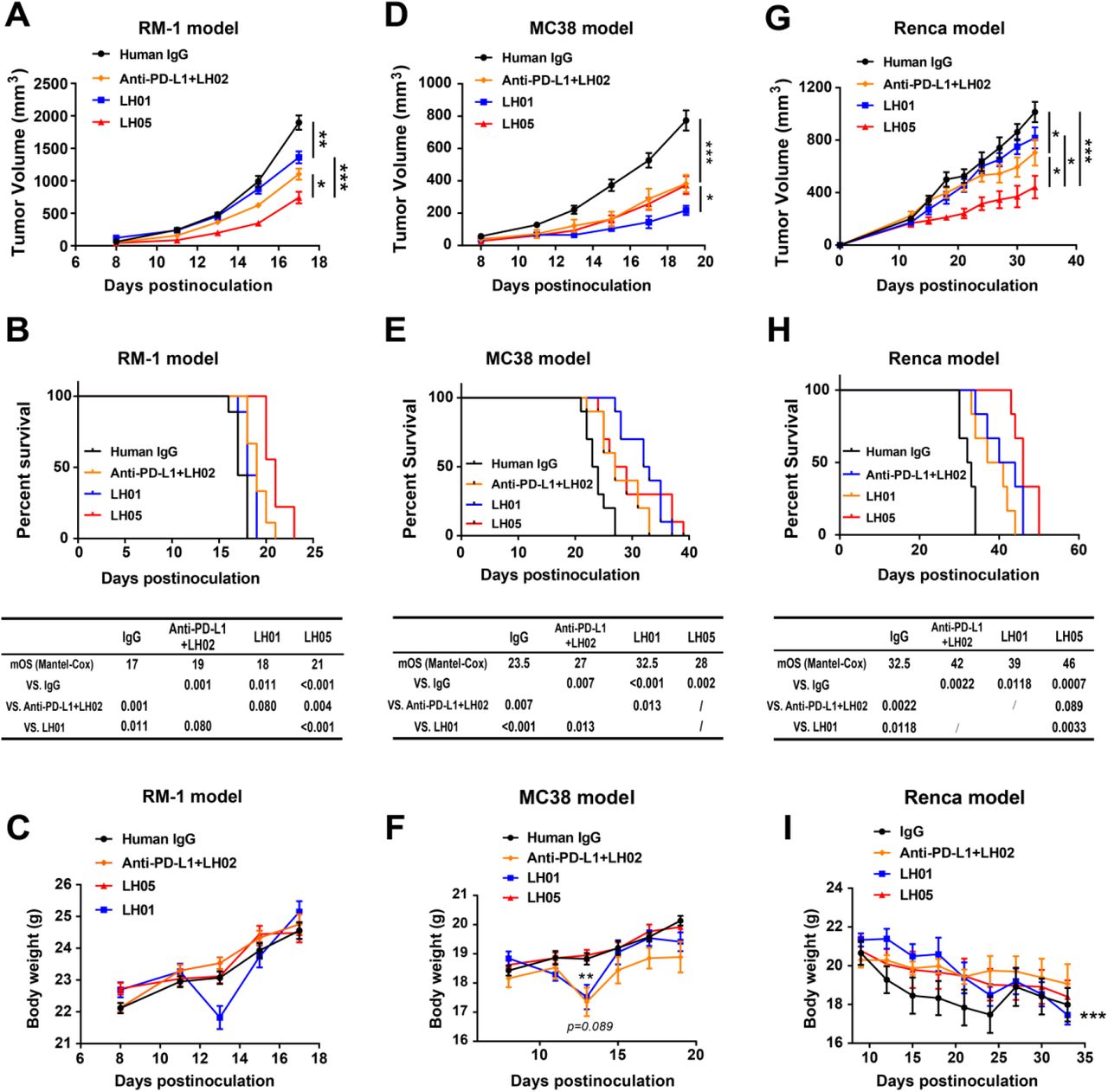
    Mice were randomized into four groups based on tumor size, with treatment initiating when tumors reached 50-100 mm 3 . Tumor growth curves were plotted over time. Mice were observed for survival starting from the day after tumor cell inoculation. The body weights of tumor-bearing mice were recorded throughout the study. (A-C) RM-1 tumor cells (5 × 10 5 ) were subcutaneously implanted into the right flank of male C57BL/6J mice. On days 9, 12, and 15 (n = 12), mice were intravenously injected with IgG control (10 mg/kg), anti-PD-L1 (10 mg/kg) + LH02 (0.25 mg/kg), LH01 (2.5 mg/kg) or LH05 (10 mg/kg). <t>(D-F)</t> <t>MC38</t> tumor cells (5 × 10 5 ) were subcutaneously implanted into the right flank of female C57BL/6J mice. On days 8, 11, 14, and 17 (n = 10), mice were intravenously injected with IgG control (10 mg/kg), anti-PD-L1 (10 mg/kg) + LH02 (0.25 mg/kg), LH01 (2.5 mg/kg) or LH05 (10 mg/kg). (G-I) <t>Renca</t> cells (5 ×10 5 ) were suspended in 50 μL PBS and an equal volume of matrigel and subcutaneously implanted into the right flank of female Balb/c mice. On days 9, 12, 16, and 21 (n = 6), mice were intravenously injected with IgG control (10 mg/kg), anti-PD-L1 (10 mg/kg) + LH02 (0.25 mg/kg), LH01 (2 mg/kg) or LH05 (10 mg/kg). All graphs show the mean ± SEM. * p < 0.05; ** p < 0.01; *** p < 0.001; ns, not significant.
    Renca Murine Renal Carcinoma Cell Line, supplied by ATCC, used in various techniques. Bioz Stars score: 96/100, based on 1 PubMed citations. ZERO BIAS - scores, article reviews, protocol conditions and more
    https://www.bioz.com/product/murine+renal+carcinoma+cell+line+renca/bio_rxiv__2023__08__02__551593-200-12-28?v=ATCC
    Average 96 stars, based on 1 article reviews
    renca murine renal carcinoma cell line - by Bioz Stars, 2026-08
    96/100 stars
      Buy from Supplier

    96
    ATCC murine renal cell carcinoma line renca
    Mice were randomized into four groups based on tumor size, with treatment initiating when tumors reached 50-100 mm 3 . Tumor growth curves were plotted over time. Mice were observed for survival starting from the day after tumor cell inoculation. The body weights of tumor-bearing mice were recorded throughout the study. (A-C) RM-1 tumor cells (5 × 10 5 ) were subcutaneously implanted into the right flank of male C57BL/6J mice. On days 9, 12, and 15 (n = 12), mice were intravenously injected with IgG control (10 mg/kg), anti-PD-L1 (10 mg/kg) + LH02 (0.25 mg/kg), LH01 (2.5 mg/kg) or LH05 (10 mg/kg). <t>(D-F)</t> <t>MC38</t> tumor cells (5 × 10 5 ) were subcutaneously implanted into the right flank of female C57BL/6J mice. On days 8, 11, 14, and 17 (n = 10), mice were intravenously injected with IgG control (10 mg/kg), anti-PD-L1 (10 mg/kg) + LH02 (0.25 mg/kg), LH01 (2.5 mg/kg) or LH05 (10 mg/kg). (G-I) <t>Renca</t> cells (5 ×10 5 ) were suspended in 50 μL PBS and an equal volume of matrigel and subcutaneously implanted into the right flank of female Balb/c mice. On days 9, 12, 16, and 21 (n = 6), mice were intravenously injected with IgG control (10 mg/kg), anti-PD-L1 (10 mg/kg) + LH02 (0.25 mg/kg), LH01 (2 mg/kg) or LH05 (10 mg/kg). All graphs show the mean ± SEM. * p < 0.05; ** p < 0.01; *** p < 0.001; ns, not significant.
    Murine Renal Cell Carcinoma Line Renca, supplied by ATCC, used in various techniques. Bioz Stars score: 96/100, based on 1 PubMed citations. ZERO BIAS - scores, article reviews, protocol conditions and more
    https://www.bioz.com/product/murine+renal+carcinoma+cell+line+renca/pm35980140-40-0-10?v=ATCC
    Average 96 stars, based on 1 article reviews
    murine renal cell carcinoma line renca - by Bioz Stars, 2026-08
    96/100 stars
      Buy from Supplier

    96
    ATCC renca murine renal cell carcinoma cell lines
    Mice were randomized into four groups based on tumor size, with treatment initiating when tumors reached 50-100 mm 3 . Tumor growth curves were plotted over time. Mice were observed for survival starting from the day after tumor cell inoculation. The body weights of tumor-bearing mice were recorded throughout the study. (A-C) RM-1 tumor cells (5 × 10 5 ) were subcutaneously implanted into the right flank of male C57BL/6J mice. On days 9, 12, and 15 (n = 12), mice were intravenously injected with IgG control (10 mg/kg), anti-PD-L1 (10 mg/kg) + LH02 (0.25 mg/kg), LH01 (2.5 mg/kg) or LH05 (10 mg/kg). <t>(D-F)</t> <t>MC38</t> tumor cells (5 × 10 5 ) were subcutaneously implanted into the right flank of female C57BL/6J mice. On days 8, 11, 14, and 17 (n = 10), mice were intravenously injected with IgG control (10 mg/kg), anti-PD-L1 (10 mg/kg) + LH02 (0.25 mg/kg), LH01 (2.5 mg/kg) or LH05 (10 mg/kg). (G-I) <t>Renca</t> cells (5 ×10 5 ) were suspended in 50 μL PBS and an equal volume of matrigel and subcutaneously implanted into the right flank of female Balb/c mice. On days 9, 12, 16, and 21 (n = 6), mice were intravenously injected with IgG control (10 mg/kg), anti-PD-L1 (10 mg/kg) + LH02 (0.25 mg/kg), LH01 (2 mg/kg) or LH05 (10 mg/kg). All graphs show the mean ± SEM. * p < 0.05; ** p < 0.01; *** p < 0.001; ns, not significant.
    Renca Murine Renal Cell Carcinoma Cell Lines, supplied by ATCC, used in various techniques. Bioz Stars score: 96/100, based on 1 PubMed citations. ZERO BIAS - scores, article reviews, protocol conditions and more
    https://www.bioz.com/product/murine+renal+carcinoma+cell+line+renca/pm35874722-196-10-21?v=ATCC
    Average 96 stars, based on 1 article reviews
    renca murine renal cell carcinoma cell lines - by Bioz Stars, 2026-08
    96/100 stars
      Buy from Supplier

    Image Search Results


    L. bacterium protects against ccRCC tumorigenesis through its supernatant (A) Experimental design: After gut microbiota depletion with ATB, RENCA-bearing mice were gavaged with L. bacterium or P. copri 5 times/week ( n = 6/group). (B and C) Tumor growth kinetics (B) and tumor weights (C) on day 24 in mice receiving L. bacterium or P. copri ( n = 6/group). (D) 16S rRNA sequencing of fecal samples from control and L. bacterium -treated mice on days 12 and 24. (E) Experimental design: RENCA-bearing mice pretreated with ATB were gavaged with L. bacterium with or without amoxicillin in drinking water ( n = 9/group). (F and G) Tumor growth curves (F) and tumor weights (G) on day 24 in the indicated groups ( n = 9/group). (H–J) Effects of L. bacterium SN on 786-O and Caki-1 cells: colony formation (H), cell viability via CCK8 assay (I), and wound healing assay (J) with 100× and 10× dilutions of SN. Scale bars: 200 μm. (K) Experimental design: ATB-pretreated RENCA-bearing mice were gavaged with L. bacterium , heat-inactivated L. bacterium , or SN ( n = 9/group). (L and M) Tumor weights in BALB/c (L) and BALB/c-nude mice (M) on day 24 ( n = 9–10/group). Data are shown as mean ± SEM. Statistical significance was determined by two-way ANOVA (B, F, and I), unpaired two-tailed t test (C, G, L, and M), and paired two-tailed t test (J). ∗ p < 0.05, ∗∗ p < 0.01, and ∗∗∗ p < 0.001. See also and .

    Journal: Cell Reports Medicine

    Article Title: Intestinal Lachnospiraceae bacterium -derived propionate inhibits the progression of clear cell renal cell carcinoma

    doi: 10.1016/j.xcrm.2025.102410

    Figure Lengend Snippet: L. bacterium protects against ccRCC tumorigenesis through its supernatant (A) Experimental design: After gut microbiota depletion with ATB, RENCA-bearing mice were gavaged with L. bacterium or P. copri 5 times/week ( n = 6/group). (B and C) Tumor growth kinetics (B) and tumor weights (C) on day 24 in mice receiving L. bacterium or P. copri ( n = 6/group). (D) 16S rRNA sequencing of fecal samples from control and L. bacterium -treated mice on days 12 and 24. (E) Experimental design: RENCA-bearing mice pretreated with ATB were gavaged with L. bacterium with or without amoxicillin in drinking water ( n = 9/group). (F and G) Tumor growth curves (F) and tumor weights (G) on day 24 in the indicated groups ( n = 9/group). (H–J) Effects of L. bacterium SN on 786-O and Caki-1 cells: colony formation (H), cell viability via CCK8 assay (I), and wound healing assay (J) with 100× and 10× dilutions of SN. Scale bars: 200 μm. (K) Experimental design: ATB-pretreated RENCA-bearing mice were gavaged with L. bacterium , heat-inactivated L. bacterium , or SN ( n = 9/group). (L and M) Tumor weights in BALB/c (L) and BALB/c-nude mice (M) on day 24 ( n = 9–10/group). Data are shown as mean ± SEM. Statistical significance was determined by two-way ANOVA (B, F, and I), unpaired two-tailed t test (C, G, L, and M), and paired two-tailed t test (J). ∗ p < 0.05, ∗∗ p < 0.01, and ∗∗∗ p < 0.001. See also and .

    Article Snippet: The human cell lines 786-O, Caki-1, and 293T, as well as the murine renal carcinoma cell line RENCA, were purchased from the American Type Culture Collection (ATCC, Manassas, VA, USA).

    Techniques: Sequencing, Control, CCK-8 Assay, Wound Healing Assay, Two Tailed Test

    Mice were randomized into four groups based on tumor size, with treatment initiating when tumors reached 50-100 mm 3 . Tumor growth curves were plotted over time. Mice were observed for survival starting from the day after tumor cell inoculation. The body weights of tumor-bearing mice were recorded throughout the study. (A-C) RM-1 tumor cells (5 × 10 5 ) were subcutaneously implanted into the right flank of male C57BL/6J mice. On days 9, 12, and 15 (n = 12), mice were intravenously injected with IgG control (10 mg/kg), anti-PD-L1 (10 mg/kg) + LH02 (0.25 mg/kg), LH01 (2.5 mg/kg) or LH05 (10 mg/kg). (D-F) MC38 tumor cells (5 × 10 5 ) were subcutaneously implanted into the right flank of female C57BL/6J mice. On days 8, 11, 14, and 17 (n = 10), mice were intravenously injected with IgG control (10 mg/kg), anti-PD-L1 (10 mg/kg) + LH02 (0.25 mg/kg), LH01 (2.5 mg/kg) or LH05 (10 mg/kg). (G-I) Renca cells (5 ×10 5 ) were suspended in 50 μL PBS and an equal volume of matrigel and subcutaneously implanted into the right flank of female Balb/c mice. On days 9, 12, 16, and 21 (n = 6), mice were intravenously injected with IgG control (10 mg/kg), anti-PD-L1 (10 mg/kg) + LH02 (0.25 mg/kg), LH01 (2 mg/kg) or LH05 (10 mg/kg). All graphs show the mean ± SEM. * p < 0.05; ** p < 0.01; *** p < 0.001; ns, not significant.

    Journal: bioRxiv

    Article Title: Next-generation anti-PD-L1/IL-15 immunocytokine elicits superior antitumor immunity in cold tumors with minimal toxicity

    doi: 10.1101/2023.08.02.551593

    Figure Lengend Snippet: Mice were randomized into four groups based on tumor size, with treatment initiating when tumors reached 50-100 mm 3 . Tumor growth curves were plotted over time. Mice were observed for survival starting from the day after tumor cell inoculation. The body weights of tumor-bearing mice were recorded throughout the study. (A-C) RM-1 tumor cells (5 × 10 5 ) were subcutaneously implanted into the right flank of male C57BL/6J mice. On days 9, 12, and 15 (n = 12), mice were intravenously injected with IgG control (10 mg/kg), anti-PD-L1 (10 mg/kg) + LH02 (0.25 mg/kg), LH01 (2.5 mg/kg) or LH05 (10 mg/kg). (D-F) MC38 tumor cells (5 × 10 5 ) were subcutaneously implanted into the right flank of female C57BL/6J mice. On days 8, 11, 14, and 17 (n = 10), mice were intravenously injected with IgG control (10 mg/kg), anti-PD-L1 (10 mg/kg) + LH02 (0.25 mg/kg), LH01 (2.5 mg/kg) or LH05 (10 mg/kg). (G-I) Renca cells (5 ×10 5 ) were suspended in 50 μL PBS and an equal volume of matrigel and subcutaneously implanted into the right flank of female Balb/c mice. On days 9, 12, 16, and 21 (n = 6), mice were intravenously injected with IgG control (10 mg/kg), anti-PD-L1 (10 mg/kg) + LH02 (0.25 mg/kg), LH01 (2 mg/kg) or LH05 (10 mg/kg). All graphs show the mean ± SEM. * p < 0.05; ** p < 0.01; *** p < 0.001; ns, not significant.

    Article Snippet: RM-1 murine prostate carcinoma cell line, MC38 murine colon carcinoma cell line, Renca murine renal carcinoma cell line, and U251 human glioblastoma cell line were obtained from the American Type Culture Collection (ATCC).

    Techniques: Injection, Control